The cysticercosis cyst structure is depicted in the images below. The diagramatic representation shows the features of the cyst used to attach onto the host's tissues. The actual structure of the tapeworm cyst, or larvae can be seen below. The scolex, sucker, and hooklets are all used to adhere to host tissue. The hooklets and sucker will be used to extract nutrients from the host tissue to allow the cyst to survive.
 |
| Source 6, Source 7. |
 |
| Panel A shows the fine structure of the cyst, and the wall tat surrounds it. Nervous tissue can be seen outside of the scolex (white arrow). Panel B shows a close view of the scolex, and the surrounding nervous tissue is disorganized, loose, and lacking in the dense structure of normal nervous tissue. Healthy nervous tissue will be densely packed with neuronal cell bodies, axons, and dendrites, as well as supportive cells such as astrocytes and oligodendrocytes. This tissue loose due to inflammation and white blood cell infiltration resulting in disorganized tissue. Panel C shows the outside wall of the cyst. Source 8 |
The previous image was taken from the case of a 53 year old woman who presented with acute back pain, and no other symptoms. MRI, surgical removal, and pathological examination revealed a cysticercal cyst. The above images show the cyst removed from the conus medullaris region of the spinal cord. This location would result in inflammation both local and regional, evidenced by the patient's "incapacitating low back pain with lower limb irradiation" (8). The tissue surrounding the cyst shows inflammation and cell necrosis. Normal healthy tissue will show none of the inflammation or cell necrosis visible in panel B of the image.
 |
| Source 9 |
 |
This image depicts a large cyst within brain parenchyma.
Source 3 |
This image shows healthy spinal cord tissue from the thoracic region. This tissue is from the medullary cone of the spinal cord. This tissue is composed of myelinated nerve fibers and multipolar nerve fibers. No inflammation or tissue separation is seen, unlike in the above panel B. This tissue is densely packed with nerve cells and supportive cells such as astrocytes and oligodendrocytes. No severe white blood cell infiltration is seen here, and there is no presence of inflammation. The tissue is densely packed and organized. Neurologic infection of cysticercosis will result in disorganization of the macrostructure of the brain, as depicted in the image to the left. This can result in symptoms as minor as headaches, and as severe as seizures, and may result in death in some cases.
 |
This image depicts a microscopic image from image 2 above. The cyst wall is depicted by the arrow, and inflamed tissue can be seen surrounding the entire cyst. Aggressive lymphocyte infiltration can be seen, along with overall tissue disorganization. The different cortical layers of the brain are not visible in this view due to the immune system response, and the cyst itself disrupting normal tissue structure.
Source 3
This image depicts a close up view of the black box in image 3A. Surrounding the cyst, the nervous tissue is filled with white blood cells, resulting in a loss of normal tissue structure. The arrows show a degenerating cell wall due to the immense immune response generated by the cyst. The lymphocyte and plasma cell flooding of the surrounding tissue has resulted in barely distinguishable nervous tissue.
Source 3.
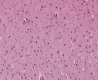
In contrast to the images above, healthy nervous tissue can be seen in this slide. The cortex of the brain is densely packed with nervous cells, and no white blood cells are easily visible here. This tissue is not inflamed, and the outer cortical cell layers of the brain are readily visible. Large pyramidal cells and smaller martinotti neurons can be identified in this tissue.
Source 10
This image shows a closer view of healthy brain tissue. Compared to the images of inflamed tissue above, this tissue is incredibly dense. Again, no leukocyte aggregation is present, therefore normal tissue function is not disrupted. Pyramidal cells, oligodendrocytes, and astrocytes are all clearly visible in this tissue.
Source 10
Images taken from a cyst found in the right frontal cortex of the brain. PAS stain. Image A is a low magnification view of the entire cyst. Images B and C are higher magnification of image A, with each scolex identified with a black arrow. Images D and E highlight the hooks of the tapeworm. Significant leukocyte migration to the tissue can be seen surrounding the cyst, especially in image D. Source 11
The hooks of the tapeworm larvae are noted by the black star. PAS stain. These are used to adhere to the host's tissue, and to gain nutrients from the host. In the top left portion and bottom right corner of the image, white blood cell infiltration can be seen. This will cause tissue inflammation and disorganization, especially as this cyst persists in the tissue for an extended duration. Source 11
H&E stain of healthy frontal brain cortex. The tissue is compact with neuronal cell bodies, axons, and glial support cells. No large spaces or leukocyte infiltration is seen, marking this as healthy tissue. Source 12
Overall, healthy tissue will not show the extreme leukocyte infiltration that is seen in the inflamed tissues containing cysts. Identifying the infected tissue is difficult due to the loss of normal identifying tissue and cellular structure. Healthy tissue will be clearly distinct, and the various cell types will be easily identifiable. Large masses and hook-looking structures are not normally found in non-infected tissues. Massive white blood cell movement is indicative of infection within a tissue and is very clearly seen histologically.
|


No comments:
Post a Comment